Active Tuberculosis Detection from Frontal Chest X-ray Images
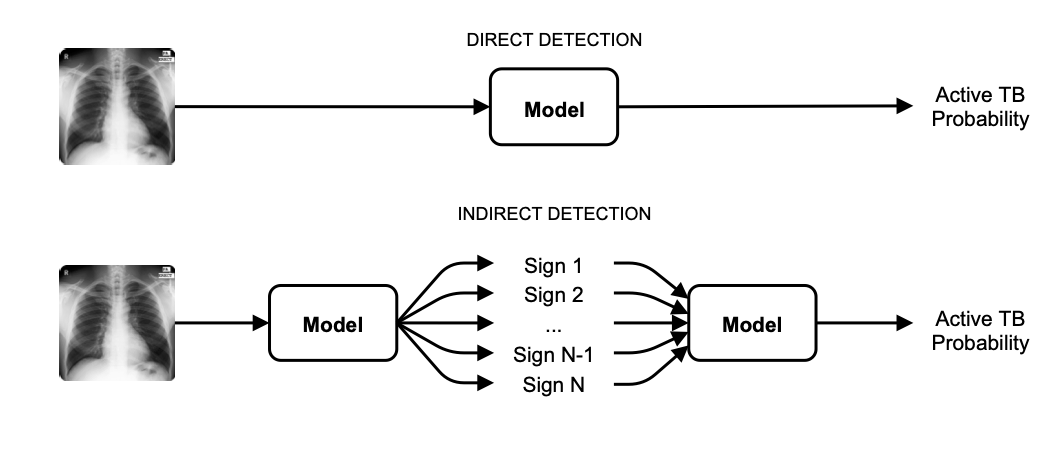
Tuberculosis remains one of the leading causes of death from a single infectious agent, and in the high-burden regions that suffer most, trained readers are scarce. The chest X-ray is therefore central to diagnosis, and in 2021 the World Health Organization accepted computer-aided detection in place of a human reader for digital images. That endorsement raised a practical question: can pulmonary tuberculosis be detected from a chest X-ray in a way that both generalises across populations and remains interpretable to the clinician who has to act on it?
Most published systems score tuberculosis directly from the pixels, an approach that tends to generalise poorly when trained on the modest amount of public tuberculosis imagery and offers little insight into its own reasoning. The hypothesis explored here was that routing the decision through the standard radiological signs a radiologist looks for would recover both properties, giving an interpretable intermediate representation that transfers better across datasets.
A model was pre-trained on tens of thousands of chest X-rays annotated with radiological signs, and a simple linear classifier then predicted the diagnosis from those signs. On three public datasets the indirect approach reached state-of-the-art performance, with test AUCs of 0.97 on Montgomery County, 0.90 on Shenzhen, and 0.93 on the Indian set, rising to 0.98, 0.98, and 0.93 when more signs were made available. An analysis of sign importance showed that it is the combination of signs, rather than any single one, that makes detection reliable.
The hypothesis is confirmed on both counts, which is the unusual part: the
interpretable route did not cost accuracy to buy transparency, it improved
generalisation as well, because the intermediate representation is one that
transfers across populations in a way raw pixel statistics do not. The answer to
the opening question is therefore yes, and the mechanism is identified — the
diagnosis should be read off the signs rather than the image. The thesis’s
clearest recommendation follows from its own bottleneck: tuberculosis datasets
should be annotated with radiological signs, since that annotation, not model
capacity, is what limits how far this reasoning can be pushed. The methods were
released through the open-source mednet library and seeded the group’s later
work on trustworthy and bias-aware tuberculosis screening.