Clinically Interpretable Computer-Aided Diagnostic Tool for Fibromuscular Dysplasia Detection
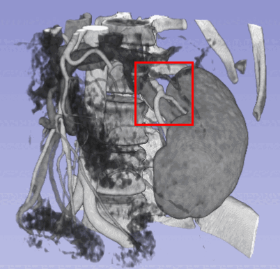
Fibromuscular dysplasia (FMD) is a rare, non-atherosclerotic vascular disease that remains hard to diagnose because its most common symptoms are non-specific, and because it most often involves the renal arteries where it is easy to overlook. That diagnostic difficulty motivated the question behind this thesis: can neural networks help detect FMD in medical imaging, and do so in a way a clinician can actually interpret?
Convolutional networks have been strikingly successful across medical image analysis, including segmentation, but a black-box detector is of little use in a clinic where a diagnosis must be justified. The hypothesis was that neural networks could detect FMD while remaining interpretable, if their decisions were exposed through saliency maps that show which regions drove them.
Four strategies for coping with a cohort of fewer than 200 patients were tried: patch-based training, contrastive pre-training, treating both kidneys as one sample, and generating one sample per kidney with non-vascular structures removed using prior anatomical knowledge. Only the last converged at all; the other three failed outright. To make the explanations testable rather than merely decorative, volumetric saliency mapping was paired with an extension of an established faithfulness score to three-dimensional models. A control experiment then exposed how narrow the margin was: a subset of control scans is never classified correctly, and reintroducing thirteen of them dropped precision from 59 to 40 percent, accuracy from 63 to 50 percent, and the area under the curve to 0.49 — indistinguishable from guessing.
The hypothesis survives only in weakened form. Neural networks can be made interpretable here, and the metric extension makes the interpretation auditable, but detection itself is not yet reliable enough for the interpretation to be worth much. The value of the answer lies in its diagnosis: because the failure tracks specific scans and the preprocessing applied to them rather than the choice of architecture, the remedy is to constrain the model anatomically — isolate the arteries and classify only those. That recommendation was taken up directly by the group’s next thesis on the disease, where it did indeed improve detection, and the gender bias flagged here remains open. The work stands as the interpretability-first foundation of the FMD collaboration with CHUV.