Automatic Grading of Vasculitis Inflammation in Fluorescein Angiography Images
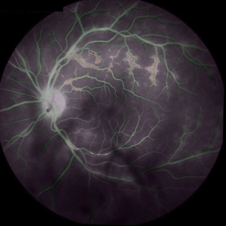
Vasculitis is an inflammatory disease that attacks the retinal blood vessels and can lead to loss of vision. It is best revealed by fluorescein angiography (FA), in which a fluorescent dye is injected into the bloodstream: vessels damaged by the disease leak part of the dye, and the resulting stain becomes visible in the images a few minutes after injection. At the Jules-Gonin Eye Hospital the severity is scored with the standard Tugal-Tuktun scale, on which retinal vascular leakage is rated focal, multifocal, or diffuse. That judgement rests on an expert eye and years of ophthalmology training, which raises the question at the heart of this thesis: can the vascular leakage that drives the grade be detected and quantified automatically from an FA exam?
Until then, automated analysis of retinal inflammation had mostly targeted diabetic retinopathy, or the global detection of hyper-fluorescence across an exam, rather than the vasculitis-specific leakage that clinicians actually grade. The difficulty, and the hypothesis this thesis set out to test, is that pathological leakage can be told apart from the vessels themselves and from the fluorescence of other retinal structures by first anchoring the analysis on the vasculature. Because the vessels are the only bright structures in the early frames of the FA time-lapse, segmenting them there and then aligning those masks onto the late frames should isolate the perivascular region where staining is expected, so that leakage appears simply as retinal background that has become abnormally bright.
The work was carried out inside the multi-centre CAD4IED project, bringing together the Jules-Gonin Eye Hospital, Idiap, the Luzern Kantonsspital, and the CHU Grenoble. A full pipeline was built, from raw clinical FA data to a leakage grade. Several image-registration strategies were compared against standard SIFT, and alignment was validated with a purpose-built parallel-view criterion, lifting overall frame-registration success from 46.5 to 66.8 percent. The gain landed where it matters: the late frames on which leakage is visible nearly doubled per study, from 1.44 to 2.86 registered frames on average, and the share of studies left with no usable late frame fell from 22.2 to 9.5 percent. Computer-vision methods then produced vessel and background masks that segmented the hyper-fluorescence on the late frames, and grading strategies mapped the outcome onto the Tugal-Tuktun categories. A study set of 543 patients was prepared for expert annotation; on an initial 60-image subset annotated by a single grader, the pipeline detected diffuse staining well enough to grade cases correctly.
The hypothesis is supported: anchoring the analysis on the vasculature does separate pathological leakage from the vessels themselves and from other fluorescent structures, and the opening question can be answered affirmatively, with the qualification that the evidence rests on a single grader and sixty images. The finding that generalises beyond this dataset is upstream of the segmentation, and was not the one the thesis set out to make: on unfiltered clinical data, registration is the binding constraint, since a leakage detector cannot be assessed at all on a study whose late frames could not be aligned, and a fifth of studies started out in that position. Getting the alignment right is therefore a precondition for trusting anything downstream of it. The results are preliminary and expected to improve once the parameters are tuned against the fully annotated set. Beyond its own numbers, the project laid the methodological groundwork for the group’s later clinic-ready uveitis scoring system, UveAI.