Beyond the Last Frame: Temporal Deep Learning for Automated Grading of Retinal Inflammation in Fluorescein Angiography
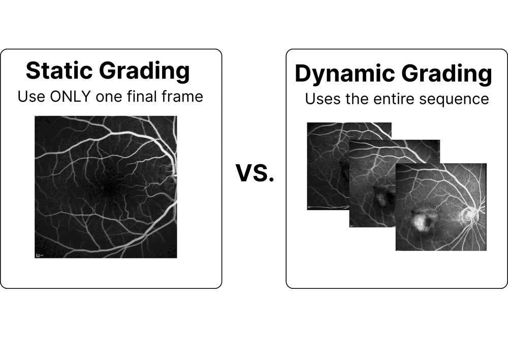
Fluorescein angiography is not a photograph but a film: a fluorescent dye is injected into the bloodstream and the camera keeps shooting for several minutes while the dye transits the retina. Clinicians separate the hyperfluorescence patterns precisely by how they behave over that interval, since leakage spreads and blurs, pooling fills a cavity with a sharp border, staining brightens without expanding, and a window defect appears early and stays geographically fixed. The group’s existing grading pipeline nonetheless collapsed each examination to a single late frame, reaching a macro ROC-AUC of 0.82 on multiclass hyperfluorescence typing while discarding the very evolution that defines several of the classes it was asked to name. The question the thesis pursued is whether reading the whole sequence recovers that discarded signal.
Earlier work in the group had established the single-frame reference this thesis had to beat, adapting retinal foundation models to angiography and settling on a compact RETFound Green Vision Transformer. What that line of work had not attempted was sequence modelling over an entire examination, and the wider literature offered temporal encoding strategies whose cost and interpretability were untested on shared research infrastructure. Two hypotheses followed. The first is that the temporal ordering of frames carries diagnostic information the last frame throws away, recoverable by passing per-frame embeddings through a lightweight recurrent model. The second, sharper hypothesis is that this only works if the image backbone is allowed to adapt to the sequence task, so that sequencing embeddings from a frozen backbone would not be enough.
Work was carried out on the public 3rd APTOS angiography benchmark, deliberately chosen over clinical data so the method would remain reproducible and publishable. The dataset carries no structured timing, so elapsed time was recovered by an OCR pipeline benchmarked against 150 hand-checked frames, reaching 96 percent accuracy and yielding 32,834 validated timestamps together with a frame eligibility filter. Phase boundaries were then calibrated from the data itself with a linear discriminant probe, landing at 103 and 518 seconds and outperforming the textbook clinical convention on every separability metric. Twelve frames, four per phase, were passed through the RETFound Green backbone into a two-layer GRU, with the existing classification head left untouched so that any gain would be attributable to the temporal block alone. With the backbone frozen, no temporal aggregation reliably beat a single well-chosen frame, a genuine negative result. Fine-tuning backbone and GRU jointly improved every graded task, raising macro ROC-AUC from 0.82 to 0.87, with the largest gains exactly where behaviour over time is diagnostic: pooling from 0.76 to 0.83 and window defect from 0.78 to 0.85. A separate controlled study found that reducing embedding dimensionality by principal component analysis before the sequence model helped nowhere, and that branch was dropped.
The evidence supports both hypotheses, and the second is the transferable one: temporal information is real, but it is only reachable by co-adapting the image backbone and the sequence model together, so bolting a recurrent layer onto frozen features buys nothing. Two limits bound the claim. The results rest on one public benchmark, and external validation on the Jules-Gonin clinical set of 543 patients was prepared and scoped but could not be run inside the internship window, because clearance for identifiable patient data takes longer than six months. Timing itself was reconstructed rather than measured, a weakness that disappears on hospital data where native timing metadata is recorded. The work left the group more than a number: the temporal pipeline was integrated into the MedNet research framework with tests and continuous integration rather than kept as a private script, its findings were accepted as an abstract at the Swiss AI in Medicine Initiative meeting in June 2026, and the clinical validation and an explicit time-embedding variant were handed over as the two next steps.