Towards Computer-Aided Diagnosis of Fibromuscular Dysplasia: Abnormality Detection in the Renal Artery Using Deep Learning
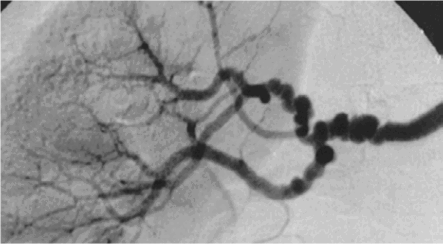
Fibromuscular dysplasia (FMD) is an under-recognised, non-atherosclerotic disease of the arteries that manifests as subtle abnormalities, from the tell-tale string-of-beads to stenosis, aneurysm, dissection, and tortuosity. Many patients stay asymptomatic until a serious event, and the signs are easy to overlook on imaging. Computed tomography angiography (CTA) is a standard way to look for them, which raised the question this thesis pursued: can deep learning detect FMD-related abnormalities in the renal arteries from CTA, to support computer-aided diagnosis?
Because the abnormalities are subtle and the disease is uncommon, automated detection is hard, and it is made harder still by the small annotated cohorts that such a rare condition affords. Earlier computer-aided work rarely targeted FMD specifically. The hypothesis was that three-dimensional convolutional networks, helped by first isolating the arteries, could flag abnormalities reliably even from a modest dataset.
Working from a cohort of 126 contrast-enhanced CTA volumes cropped to the renal region, three progressively more informed pipelines were compared under five-fold cross-validation: a baseline 3D convolutional network trained from scratch, the same network fed only presegmented vessels, and a frozen vascular foundation model used as a feature extractor. The baseline reached an area under the curve of 0.849 ± 0.018. Presegmenting the vasculature raised this to 0.900, with average precision improving from 0.774 to 0.875 and accuracy from 0.746 to 0.831. The foundation model, by contrast, matched the baseline at 0.851 and did not approach the segmentation-based pipeline. Consistent mild overfitting was observed throughout, and with only five folds most differences fall short of statistical significance.
The hypothesis is supported in direction but not yet in strength. Deep learning does detect FMD-related abnormalities from modest cohorts, and isolating the arteries first is what makes the difference — the gain is best explained as anatomical inductive bias, since restricting the field of view removes the contextual confounders a small dataset invites a model to exploit. The negative half of the answer is equally useful: a pretrained vascular foundation model brought no benefit over training from scratch, so supplying vessel information directly beat inheriting it. The honest verdict is a promising direction on evidence too thin to be conclusive, with lesion-level annotation identified as the prerequisite for the next step.