Ophthalmology — Uveitis
AI-assisted grading of intraocular inflammation in uveitis from retinal fluorescein angiography, developed with the Jules-Gonin Eye Hospital.
Partnerships
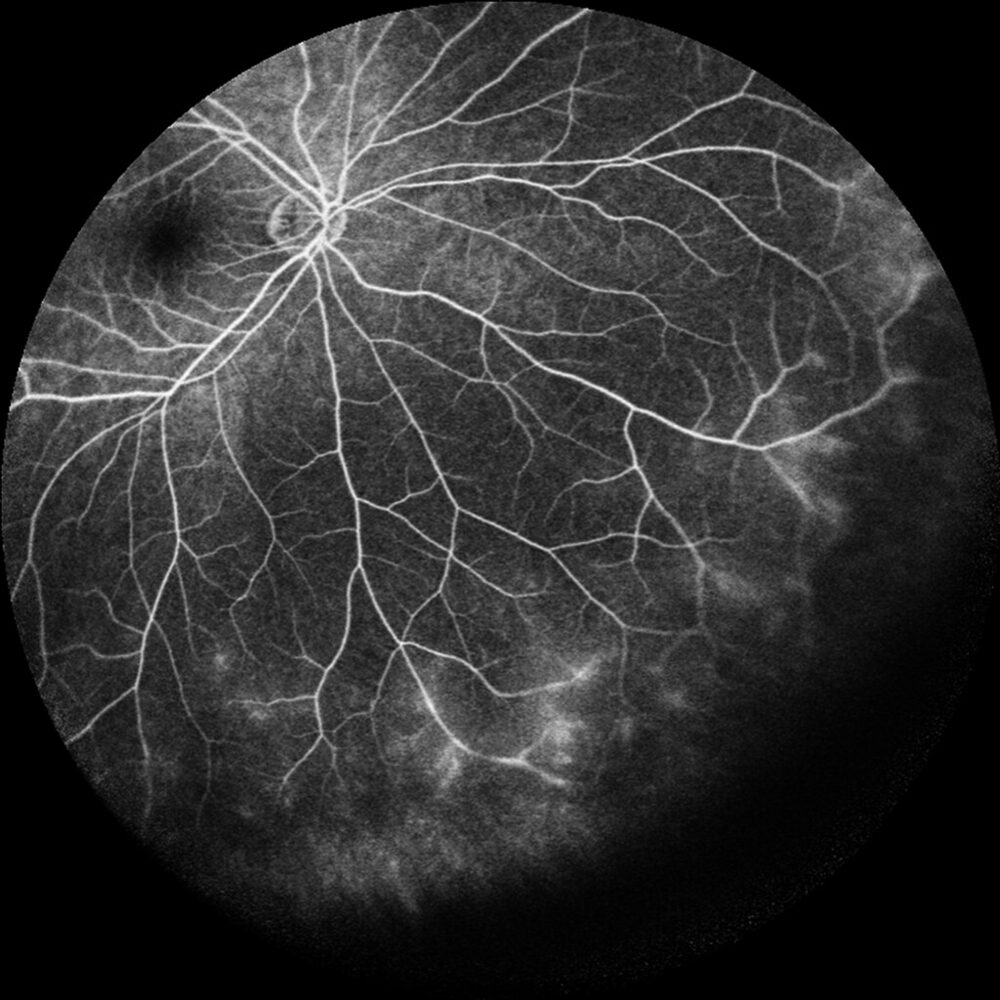
Cover: ultra-widefield fluorescein angiography of anterior uveitis showing peripheral vascular leakage, from Chi et al., PLOS ONE 2015, used under CC BY 4.0.
Uveitis is a leading cause of preventable blindness, and treating it well depends on grading intraocular inflammation reliably. That inflammation is read from widefield fluorescein angiography, a task that is slow and on which experienced clinicians often disagree. With the Jules-Gonin Eye Hospital (HOJG) and partner clinics, my group builds models that grade retinal inflammatory signs directly from angiography, aiming at scoring that is reproducible, interpretable, and ready for the clinic.
Major achievements
The work advanced from grading a single inflammatory sign to scoring the retina comprehensively. We first showed that retinal vasculitis can be graded fully automatically from angiographic time-lapses drawn straight from hospital imaging devices, with no manual curation of the images: on 3,205 images from 242 eyes the pipeline reached an F1 of 0.81 and an area under the curve of 0.86, against 0.57 and 0.66 for an intensity-based baseline. Moving from detection to grading was the harder problem, because clinical severity is recorded on ordered categories in which confusing adjacent grades matters far less than confusing the extremes. Training transformer models against that ordinal structure, and scoring them with a measure that respects it, we graded four signs of the posterior pole — vascular and capillary leakage, macular oedema, and optic disc hyperfluorescence — across 40,987 images from 1,042 eyes. The models matched expert graders sign by sign and, averaged over signs, marginally exceeded the agreement observed between the human experts themselves.
These components were assembled into UveAI, an end-to-end system combining six specialised models, four for the posterior pole and two for the periphery, into one inflammation score aligned with the semi-quantitative scale already used in the clinic. Trained on 3,220 images from 644 eyes and tested against three further uveitis specialists from different hospitals, its composite score tracked the reference grader at a correlation of 0.96, above the 0.84 observed between the human experts, with a mean area under the curve of 0.952 across the six signs; saliency analysis confirmed the models attend to clinically meaningful structures rather than to acquisition artefacts. The work leaves behind the largest annotated angiography dataset in uveitis to date and the first automated scoring to span both the posterior pole and the periphery, with the honest caveats that validation is so far single-centre, single-device, and anchored on one senior grader. A parallel study of foundation models tempered hopes of easy reuse: pretraining on a related eye modality helps only when the self-supervised objective preserves representational diversity, and can otherwise collapse the model’s attention and actively harm transfer.
Key publications
- Beyond the Last Frame: Temporal Modelling of Fluorescein Angiography for Hyperfluorescence Classification. Ophthalmic Medical Image Analysis (OMIA), MICCAI 2026 Workshops, 2026. Conference Paper · PDF · Software
- UveAI: clinic-ready scoring of retinal inflammation in uveitis on widefield fluorescein angiography using AI. Scientific reports, 2026. Journal Article · doi:10.1038/s41598-026-46069-w · Software
- When Specialization Helps (and Hurts): Cross-Modality Transfer in Ophthalmic Imaging with Foundation Models. 2026 IEEE 23rd International Symposium on Biomedical Imaging, 2026. Conference Paper · doi:10.1109/isbi61048.2026.11515636 · Homepage · Software
- Automatic transformer-based grading of multiple retinal inflammatory signs in uveitis on fluorescein angiography. Computers in Biology and Medicine, 2025. Journal Article · doi:10.1016/j.compbiomed.2025.110327 · PDF · Related Preprint doi:10.2139/ssrn.4960069
- Fully Automatic Grading of Retinal Vasculitis on Fluorescein Angiography Time-lapse from Rheumatologic Uveitis. 2023 IEEE 36th International Symposium on Computer-Based Medical Systems (CBMS), 2023. Conference Paper · doi:10.1109/cbms58004.2023.00301
Software
- mednet. A framework for developing and analysing deep neural network architectures for medical data (2D/3D images and signals), with ready access to many public medical-imaging datasets and extensible model architectures. Software GPL-3.0 · Docs · PyPI · conda-forge · Source
Supervised theses
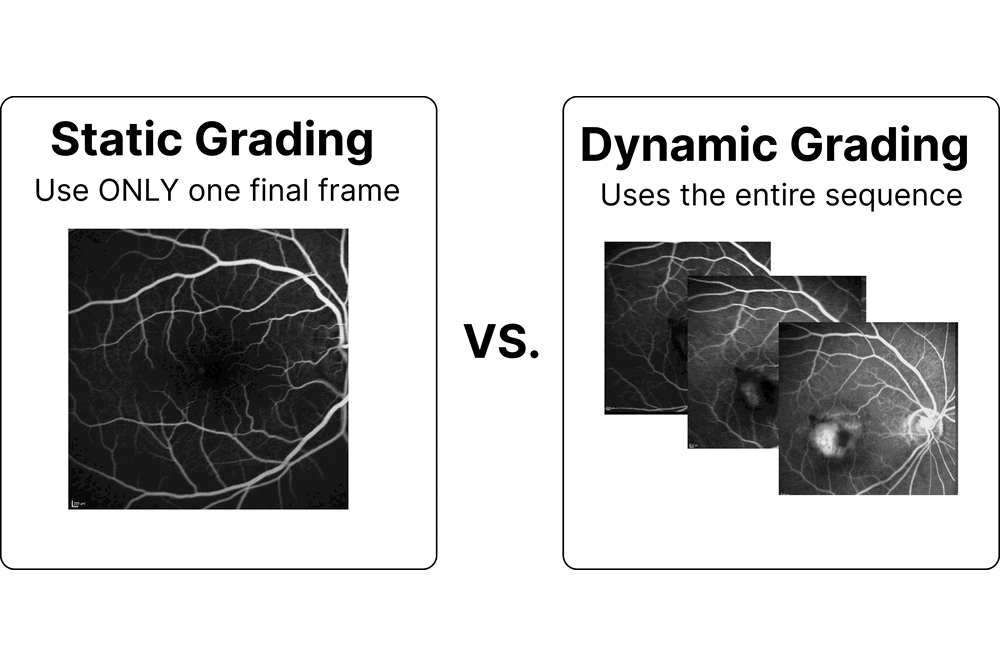
Beyond the Last Frame: Temporal Deep Learning for Automated Grading of Retinal Inflammation in Fluorescein Angiography
Fluorescein angiography films the eye for several minutes, yet the group's grading model was reading only the final photograph. This thesis taught it to watch the whole sequence, lifting grading accuracy where the disease is defined by how it changes over time.
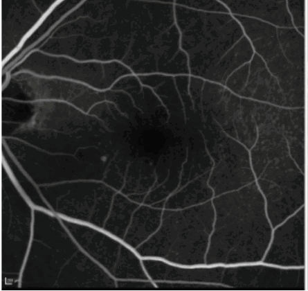
Evaluating Cross-Domain Adaptation Strategies for Foundation Models: A Study on Fluorescein Angiography
Fluorescein angiography reveals retinal disease but is far less annotated than ordinary fundus photography. This thesis tested whether AI "foundation models" trained on fundus images can be adapted to angiography efficiently, including for grading uveitis inflammation with the Jules-Gonin Eye Hospital.
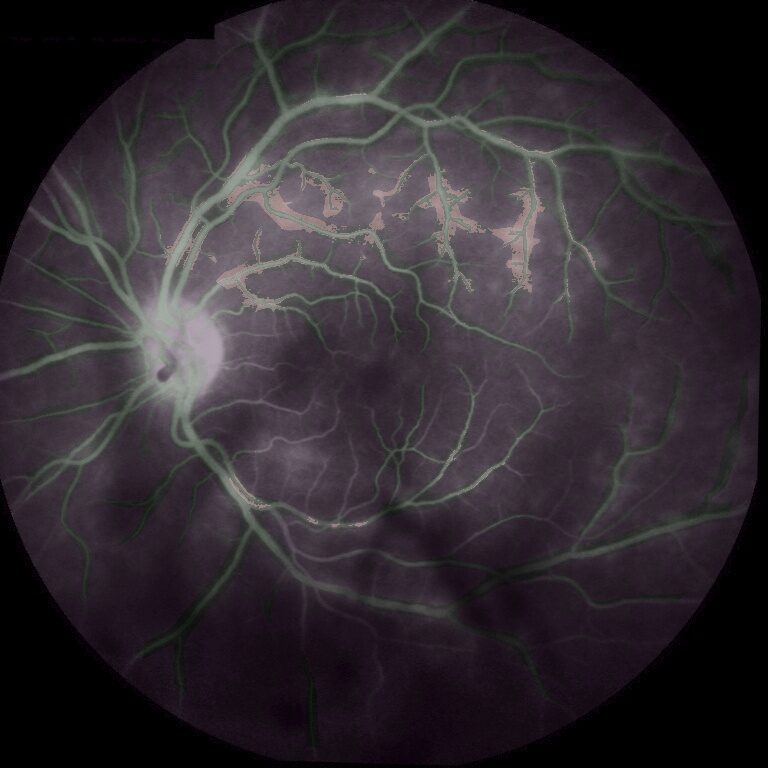
Automatic Grading of Vasculitis Inflammation in Fluorescein Angiography Images
Vasculitis inflames the blood vessels at the back of the eye and can cost a patient their sight. This thesis built an automatic pipeline that detects and grades the dye leakage from angiography, tested on 543 patients at Jules-Gonin.