Radiology — Robust Image Analysis
Interpretable, generalisable computer-aided analysis of radiological images — detecting tuberculosis on chest X-rays and extracting stable quantitative biomarkers from CT — built to hold up across scanners and populations.
Partnerships
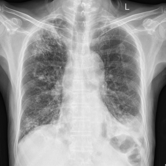
A radiological image, a chest X-ray or a CT slice, is only as useful as the information a computer can reliably pull from it, and that reliability is exactly what tends to break when the scanner, the site, or the patient population changes. This work builds computer-aided analysis of radiological images that stays interpretable, fair, and stable enough to trust outside the lab, from detecting disease to quantifying it.
Major achievements
For tuberculosis, where high-burden regions have too few trained readers and opaque models fall apart on unfamiliar images, we route the decision through the radiological signs a radiologist would name and predict the diagnosis from those, reaching areas under the curve of about 0.97 on Montgomery, 0.90 on Shenzhen and 0.93 on the Indian set while keeping the reasoning legible. We then pressed the harder question of whether a model is right for the right reasons, and the answer was sobering: classifiers scoring a perfect area under the curve on the standard benchmark fell to 0.79 on an unseen external cohort, and their saliency maps concentrated outside the lungs entirely. Pre-training on a large, only loosely related radiograph collection and balancing the classes through the loss recovered both properties at once, lifting external performance to 0.88 while roughly quadrupling the overlap between model attention and expert-drawn disease regions. And because much of the world still records chest X-rays on film, we measured what photographing those films costs a lung-segmentation model: its precision-recall area falls from 0.99 on digital images to 0.90 on digitised ones, a gap any field deployment must budget for.
The same fragility haunts quantitative imaging. Radiomics promises to turn a scan into
biomarkers, yet many classic features change value the moment the acquisition settings do.
Imaging a 3D-printed anthropomorphic phantom containing four classes of liver tissue and
lesion under eight acquisition protocols, thirty repeat scans each, let us score stability and
discriminative power on the same footing. Three-dimensional deep features proved at least
twice as stable across scanner variation as the steadiest hand-crafted family, and some three
times as stable as hand-crafted features overall, while giving up almost nothing in their
ability to separate the tissue classes — and this from a network trained on an unrelated
anatomical task on entirely different data. Wavelet features were the most discriminative of
all but the least stable, which is exactly the trade-off a biomarker study needs to see before
committing to it. Across both strands the throughline is the same, and the tooling is shared
through the open-source mednet library: for computer-aided radiology to help real clinics,
what it extracts from an image has to be interpretable, fair, and robust to the scanners and
populations it will actually meet.
Key publications
- Refining Tuberculosis Detection in CXR Imaging: Addressing Bias in Deep Neural Networks via Interpretability. 2024 12th European Workshop on Visual Information Processing (EUVIP), 2024. Conference Paper · doi:10.1109/euvip61797.2024.10772829 · Homepage · PDF · Software
- Comparing Stability and Discriminatory Power of Hand-crafted Versus Deep Radiomics: A 3D-Printed Anthropomorphic Phantom Study. 2024 12th European Workshop on Visual Information Processing (EUVIP), 2024. Conference Paper · doi:10.1109/euvip61797.2024.10772813
- Pulmonary Tuberculosis Screening from Radiological Signs on Chest X-Ray Images Using Deep Models. Union World Conference on Lung Health, 2022. Presentation
- Development of a lung segmentation algorithm for analog imaged chest X-Ray: preliminary results. Anais do 15. Congresso Brasileiro de Inteligência Computacional, 2021. Conference Paper · doi:10.21528/cbic2021-123 · PDF
Software
- mednet. A framework for developing and analysing deep neural network architectures for medical data (2D/3D images and signals), with ready access to many public medical-imaging datasets and extensible model architectures. Software GPL-3.0 · Docs · PyPI · conda-forge · Source
Supervised theses
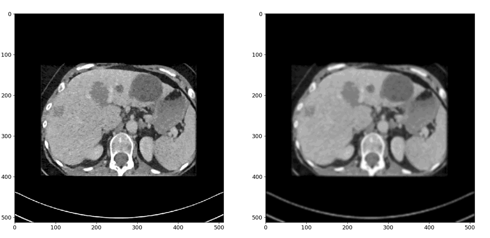
Stability and Accuracy of Radiomic Features in Medical Image Classification
A radiomic biomarker is only useful if it does not change every time the scanner settings do. This thesis looked for image features that stay both stable and informative across acquisition protocols.
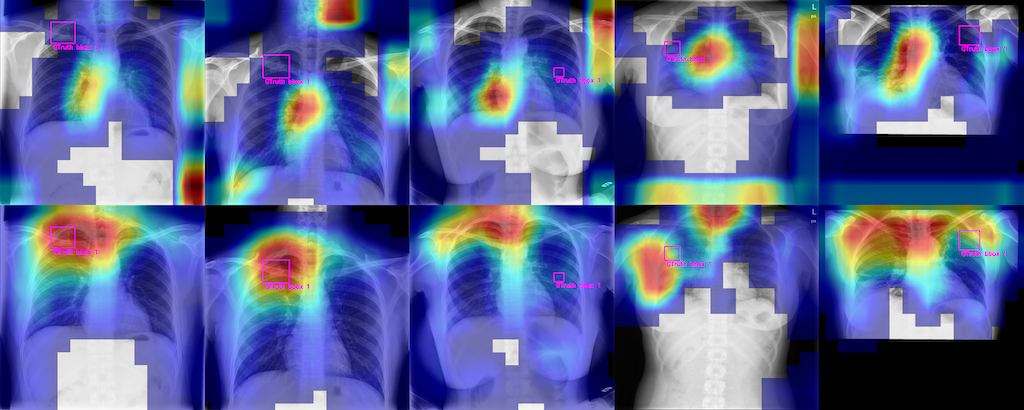
Explaining CNN-Based Active Tuberculosis Detection through Saliency Mapping
Deep networks can flag active tuberculosis on chest X-rays, but can we trust the heatmaps that explain them? This thesis measured which explanation methods are actually faithful, and improved detection with more annotated data.
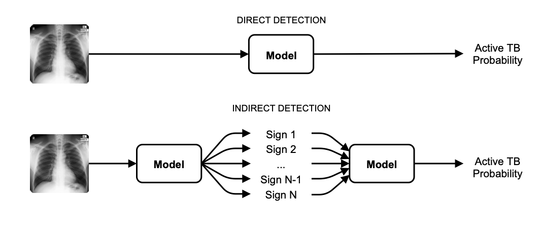
Active Tuberculosis Detection from Frontal Chest X-ray Images
Tuberculosis is often diagnosed from chest X-rays where specialists are scarce. This thesis built an interpretable, generalisable way to detect it by reading radiological signs rather than scoring the image directly.