Ophthalmology — Retinal Image Analysis
Segmentation and analysis of retinal fundus images (vessels, optic disc, blood flow) with minimalistic, reproducible, and clinically deployable models.
Partnerships
Retinal fundus imaging is a cheap, non-invasive window onto vascular and neurological health, and many sight-threatening diseases develop before symptoms appear. Analysing the retinal vasculature automatically therefore supports earlier and more interpretable diagnosis. A recurring principle in this work is that the models should stay small and rigorously reproducible: state of the art, but light enough to train honestly and to run in a clinic.
Major achievements
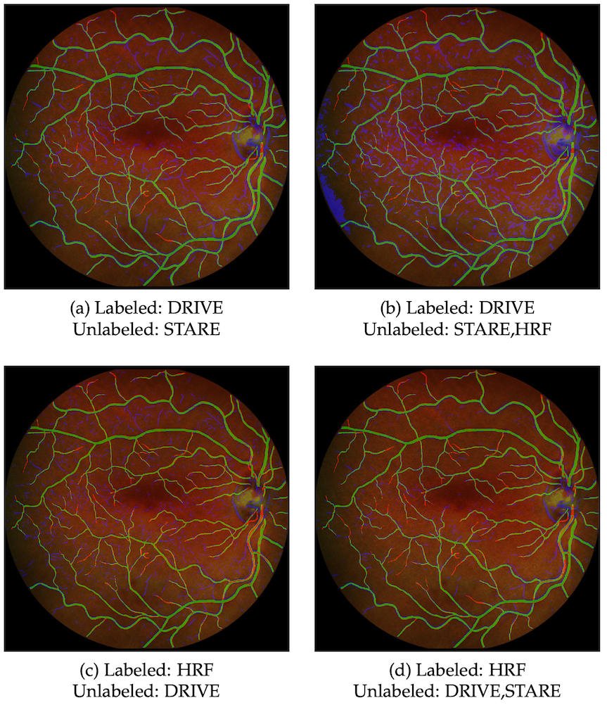
On vessel segmentation we deliberately stepped back from ever-larger networks. Collating twenty published methods, we showed that a carefully trained, minimalistic U-Net of some 34,000 parameters — one to three orders of magnitude smaller than the architectures it was competing with — closely matches the best of them, and that a simple cascaded extension, W-Net, at roughly 68,000 parameters surpasses them on the standard benchmarks. Making that claim safely meant first repairing how the field measures itself: we adopted a strict protocol that computes metrics at native resolution and fixes the decision threshold from training data alone, and a companion study showed that once scores are computed consistently the differences between most published architectures are not statistically meaningful, that high-resolution images need nothing more elaborate than rescaling, and that the entire pipeline can be shipped as open code others can rerun. The same effort produced the most comprehensive cross-dataset analysis to date, across ten databases, which made plain that segmentation is far from solved once test images differ from the training set; a simple self-labelling scheme recovers part of the lost generalisation.
Structure was only half the question. With the Grenoble team we contributed the vessel
segmentation behind an adaptive-optics laser-Doppler velocimeter that measures absolute
retinal blood flow non-invasively. Replacing conventional vessel detection with a learned
segmentation raised the share of the 12,320 adaptive-optics images yielding an automatic
calibre measurement from 64.9% to 99.5%, which is what made the clinical study tractable:
across fifteen healthy subjects total venous flow was 37.8 ± 6.8 µl/min, whereas in eyes
with retinal vein occlusion flow through the occluded vein fell to 3.51 ± 2.25 µl/min and
the tight relationship between vessel calibre and flow seen in healthy eyes largely broke
down. The thread across the project is restraint and reproducibility: the segmentation and
evaluation methods live on in the open-source mednet library, and the recurring finding,
that small models rigorously benchmarked rival heavy ones yet still generalise poorly out
of distribution, has shaped how we build and report clinically deployable retinal models
rather than chase leaderboard positions.
Experiments use public retinal benchmarks — DRIVE, STARE, CHASE-DB1, HRF, and IOSTAR.
Key publications
- Absolute retinal blood flow in healthy eyes and in eyes with retinal vein occlusion. Microvascular Research, 2024. Journal Article · doi:10.1016/j.mvr.2023.104648
- State-of-the-art retinal vessel segmentation with minimalistic models. Scientific Reports, 2022. Journal Article · doi:10.1038/s41598-022-09675-y · PDF
- On the Evaluation and Real-World Usage Scenarios of Deep Vessel Segmentation for Retinography. arXiv, 2020. Preprint · doi:10.48550/arxiv.1909.03856
Software
- mednet. A framework for developing and analysing deep neural network architectures for medical data (2D/3D images and signals), with ready access to many public medical-imaging datasets and extensible model architectures. Software GPL-3.0 · Docs · PyPI · conda-forge · Source
Supervised theses
Semantic Segmentation of Weakly Labeled Retinal Images
Labelling retinal images pixel by pixel is slow and expensive. This thesis learned to segment retinal vessels while leaning on unlabelled images, so good results need far fewer annotations.
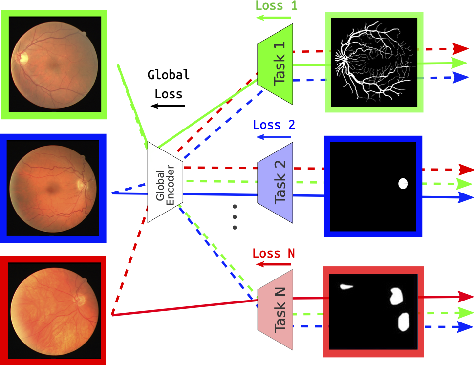
Computer-Aided Screening for Eye Diseases from 2-D Retinography
Many blinding eye diseases show no early symptoms. This thesis worked toward interpretable screening from ordinary retinal photographs by teaching a single model to segment the fundus structures that disease affects most.