Angiology — Fibromuscular Dysplasia
Machine learning for the characterisation and diagnosis of fibromuscular dysplasia, in collaboration with CHUV's angiology department.
Partnerships
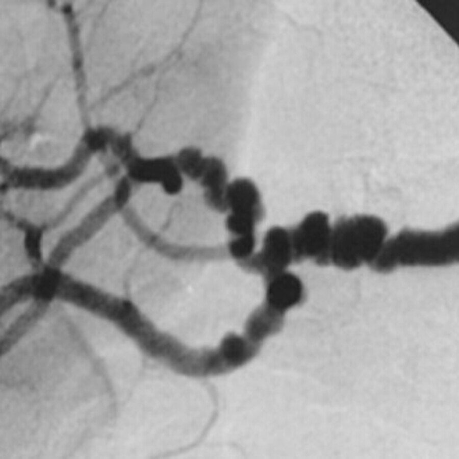
Cover: the diagnostic “string-of-beads” renal angiogram of medial fibromuscular dysplasia, from Plouin et al., Orphanet Journal of Rare Diseases 2007;2:28 (CC BY 2.0).
Fibromuscular dysplasia (FMD) is an under-recognised arterial disease that is not caused by atherosclerosis. It can lead to stenosis, aneurysm, and dissection, most often in younger women, and because it is easy to miss, the diagnosis is frequently delayed. Together with the angiology department at CHUV, we are developing machine-learning methods to help characterise and diagnose it.
Major achievements
The collaboration’s first phase asked whether three-dimensional convolutional networks can detect FMD at all from contrast-enhanced angiography, and answered with useful caution. Working from a cohort of fewer than 200 patients, we found that most standard remedies for scarce data — patch-based training, contrastive pre-training, and treating both kidneys as a single sample — failed to converge at all. Only generating one sample per kidney and stripping away non-vascular structures using prior anatomical knowledge produced a model with genuine signal. Because a diagnosis has to be justifiable, we paired the classifier with volumetric saliency mapping and extended an established faithfulness measure to three dimensions, so the explanations could themselves be tested rather than taken on trust. A control experiment showed how thin the margin was: adding a set of persistently misclassified control scans pushed performance back to chance, which located the difficulty in the data and the preprocessing rather than in the architecture.
That diagnosis pointed straight at the next step, and it proved correct. Restricting the model to the renal arteries — segmenting the vasculature first and classifying only what remains — raised the area under the curve from 0.849, for a three-dimensional network trained from scratch on the whole region, to 0.900 across 126 angiography volumes under five-fold cross-validation, with average precision improving from 0.774 to 0.875. Anatomical priors, in other words, do work that would otherwise demand far more data. A vascular foundation model used as a frozen feature extractor brought no improvement over training from scratch, a useful negative result at a moment when such models are widely assumed to help. The project remains at an early stage, and we say so plainly: the cohorts are small enough that most of these differences fall short of statistical significance, and lesion-level annotation is the obvious prerequisite for the next advance.
Supervised theses
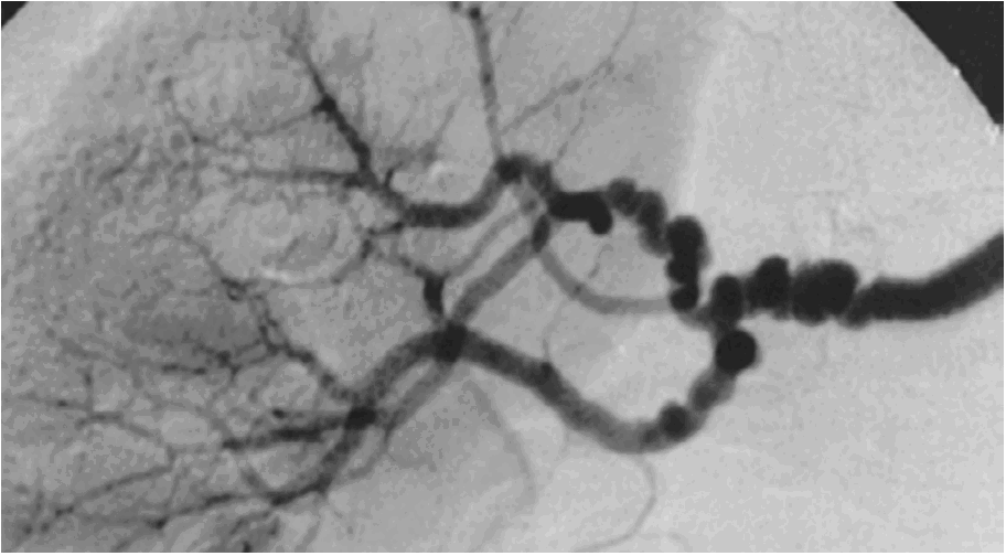
Towards Computer-Aided Diagnosis of Fibromuscular Dysplasia: Abnormality Detection in the Renal Artery Using Deep Learning
Fibromuscular dysplasia shows up as subtle abnormalities in the arteries and is easily missed. This thesis trained deep-learning models to detect them in renal-artery CT angiography, from a cohort of 126 scans.
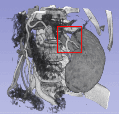
Clinically Interpretable Computer-Aided Diagnostic Tool for Fibromuscular Dysplasia Detection
Fibromuscular dysplasia is rare and easy to miss. This thesis explored neural networks that detect it in medical imaging while explaining their decisions, so a clinician can see why.